-
Address: Suite 4 (level 1), 52 Hatherley Parade, Winthrop, WA 6150.
-
Phone: 08 9332 2455
-
Email: reception@waurology.com.au

Hematuria Investigation
What Is Hematuria?
Hematuria is the presence of red blood cells in the urine.
Hematuria can be either:
- Microscopic – not visible to the naked eye
- Macroscopic – visible to the naked eye
Microscopic Hematuria
Microscopic hematuria occurs in 2-5% of people in the community. It can be normal to lose red blood cells in the urine, but not enough to show up in tests. Microscopic hematuria is defined as when a urine sample is seen under a microscope there are more than 3 red blood cells per high power field view in 2 out of 3 urine specimens.
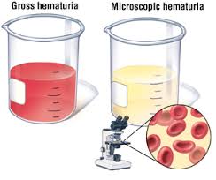 Macroscopic Hematuria
This occurs when there is more than 1ml of blood in urine. However, not all red coloured urine is due to blood. Urine can also appear red:
- After eating beetroot due to its pigment
- With medications (eg: rifampicin)
- With filtered breakdown products of muscle
Causes Of Hematuria
There are many possible causes of hematuria ranging from the trivial to the lethal. Some of the more common causes include:
- Urinary tract infection – normally in this case there is pain with urinating along with the blood in the urine.
- Stones – in the kidney or bladder
- Benign prostatic hyperplasia
- Cancers of the kidney, prostate or bladder
- Vigorous exercise
- Idiopathic (no cause found)
Diagnosis
Regardless of the type of hematuria, persistent hematuria requires investigation in order to exclude any possible serious underlying causes such as cancer.
Macroscopic hematuria
About 20% of patients with macroscopic hematuria have underlying cancer as the cause. Tests performed are:
- Imaging with a CT IVP. This is where X-ray dye is injected into the veins. The dye is then taken up by the kidneys and excreted into the ureters. As a result any abnormality of the kidneys or ureters is readily seen.
- Cystoscopy is done to assess for any possible bladder problems.
Microscopic hematuria
About 2 to 3% of patients with microscopic hematuria have underlying cancer as the cause. While CT IVP can be done in this setting an alternative is:
- Imaging with ultrasound of the kidneys to check for any causes.
- Urine cytology. This is where the urine is tested for any cancer cells shed by a possible underlying cancer. However, urine cytology is not very sensitive for low grade cancers. As such a negative result does not mean that cancer is not present.
- Cystoscopy – is required to assess the bladder.
To find out more about Flexible Cystoscopy (Male) Click Here
To find out more about Flexible Cystoscopy (Female) Click Here
Treatment
Treatment depends on cause identified and if a cause is found it is dealt with appropriately. If no cause is found further treatment is not required.